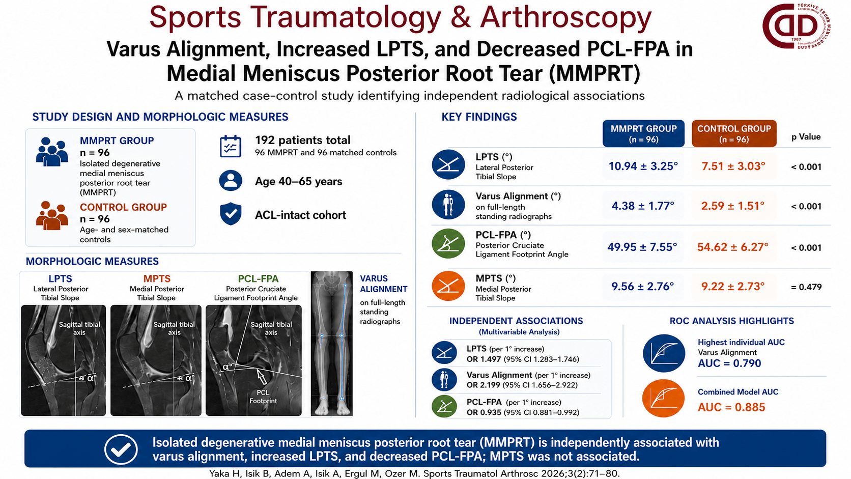
Abstract
Objective: The purpose of this study was to investigate the relationship between isolated degenerative medial meniscus posterior root tears (MMPRTs) and varus alignment, lateral and medial posterior tibial slopes (LPTS and MPTS), and the PCL tibial footprint angle (PCL-FPA).
Materials and Methods: This retrospective matched case-control study included 192 patients aged 40–65 years without ACL pathology. Ninety-six patients with MMPRT were matched 1:1 by age and sex with 96 controls, yielding 96 matched pairs. LPTS and MPTS were measured on sagittal MRI using the Hudek technique; PCL-FPA was defined as the angle between the tibial longitudinal axis and the PCL footprint line on MRI; and varus alignment was assessed by measuring the mechanical axis angle on full-length weight-bearing radiographs. Mann-Whitney U and chi-square tests were used for between-group comparisons, and Firth-penalized logistic regression analysis was used to identify independently associated parameters.
Results: Compared with the control group, the MMPRT group demonstrated significantly higher LPTS (10.94±3.25° vs. 7.51±3.03°; p<0.001) and varus alignment (4.38±1.77° vs. 2.59±1.51°; p<0.001), and significantly lower PCL-FPA (49.95±7.55° vs. 54.62±6.27°; p<0.001). MPTS was similar between the groups (p=0.479). On multivariable analysis, LPTS (OR=1.497; 95% CI 1.283–1.746; p<0.001), varus alignment (OR=2.199; 95% CI 1.656–2.922; p<0.001), and PCL-FPA (OR=0.935; 95% CI 0.881–0.992; p=0.026) were independently associated with isolated degenerative MMPRT. ROC analysis revealed the highest individual AUC for varus alignment (AUC=0.790; 95% CI 0.724–0.851), while the combined model yielded an AUC of 0.885 (95% CI 0.835–0.929).
Conclusion: Isolated degenerative MMPRT is independently associated with varus alignment, increased LPTS, and decreased PCL-FPA. MPTS was not significantly associated with MMPRT in this ACL-intact cohort. Increased LPTS may reflect altered rotational knee biomechanics related to MMPRT, whereas decreased PCL-FPA may represent a distinct posterior tibial attachment geometry related to the medial meniscus posterior root. Further biomechanical studies and external validation are needed to confirm these findings.
Graphical Abstract