Abstract
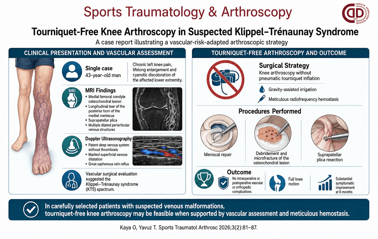
Klippel–Trénaunay syndrome (KTS) and related venous malformation disorders may complicate lower-extremity surgery because of abnormal venous circulation and the potential risks of bleeding and thromboembolic events. We report the case of a 43-year-old man with chronic left knee pain and lifelong enlargement and cyanotic discoloration of the affected lower extremity. Magnetic resonance imaging demonstrated a medial femoral condyle osteochondral lesion, a longitudinal tear of the posterior horn of the medial meniscus, a suprapatellar plica, and multiple dilated periarticular venous structures. Doppler ultrasonography showed a patent deep venous system without thrombosis but marked superficial venous dilatation and great saphenous vein reflux. Following vascular surgical evaluation, the clinical findings were considered suggestive of the KTS spectrum. Knee arthroscopy was therefore performed without pneumatic tourniquet inflation. Adequate visualization was maintained using gravity-assisted irrigation and meticulous radiofrequency hemostasis. The medial meniscal tear was repaired, the osteochondral lesion was treated with debridement and microfracture, and the suprapatellar plica was resected. No intraoperative or postoperative vascular or orthopedic complications occurred. At 6 months, the patient had regained full knee motion with substantial symptomatic improvement. This case suggests that tourniquet-free knee arthroscopy may be a feasible option for carefully selected patients with suspected venous malformations, provided appropriate preoperative vascular assessment and meticulous intraoperative hemostasis are employed.
Graphical Abstract